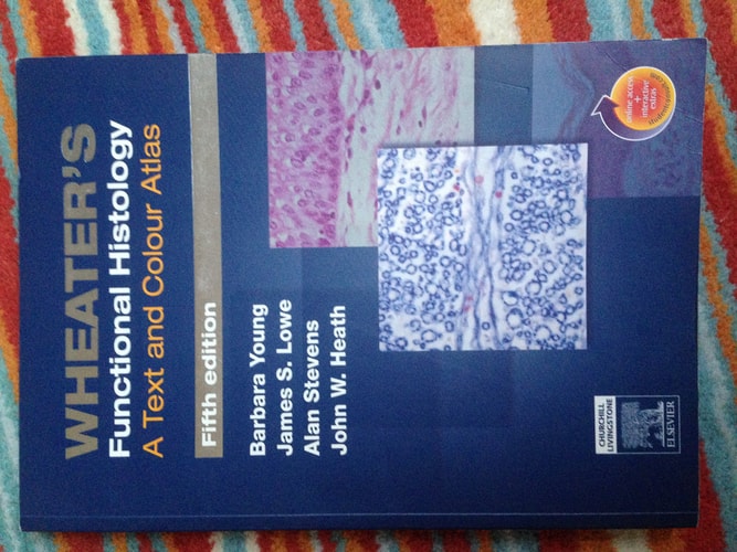
Wheater's Functional Histology: A Text and Colour Atlas
0
0
As an Amazon Associate and affiliate partner, Menrva Books earns from qualifying purchases. Learn more
Author Description
This best-selling atlas contains over 900 images and illustrations to help you learn and review the microstructure of human tissues. The book starts with a section on general cell structure and replication. Basic tissue types are covered in the following section, and the third section presents the microstructures of each of the major body systems. The highest -quality color light micrographs and electron micrograph images are accompanied by concise text and captions which explain the appearance, function, and clinical significance of each image. The accompanying website lets you view all the images from the atlas with a "virtual microscope", allowing you to view the image at a variety of pre-set magnifications. Includes access to website containing book images and additional material, extra illustrations, self tests, and more. Utilizes "virtual microscope" function on the website, allowing you to see images first in low-powered and then in high powered magnification. Incorporates new information on histology of bone marrow, male reproductive system, respiratory system, pancreas, blood, cartilage, muscle types, staining methods, and more. Uses Color coding at the side of each page to make it easier to access information quickly and efficiently. Includes access to www.studentconsult.com - where you'll find the complete text and illustrations of the book online, fully searchable · "Integration Links" to bonus content in other STUDENT CONSULT titles · 300 new USMLE-style review questions, with answers and rationales · content clipping for handheld devices · an interactive community center with a wealth of additional resources · and much more!
Menrva's Summary
THE THESIS
This atlas provides a comprehensive visual and textual guide to the microstructure of human tissues. It begins with fundamental concepts of cell structure and replication, progresses to the basic tissue types, and then details the microstructures of the major body systems. The content is enhanced by high-quality color light and electron micrographs, accompanied by concise explanations of appearance, function, and clinical significance.
THE SCOPE MAP
- Detailed coverage of general cell structure and replication.
- Explanation of basic tissue types.
- Microscopic anatomy of major body systems.
- High-quality color light and electron micrographs.
- Concise text and captions explaining appearance, function, and clinical significance.
- New information on histology of bone marrow, male reproductive system, respiratory system, pancreas, blood, cartilage, and muscle types.
- Color coding for efficient information access.
- Access to an accompanying website with a 'virtual microscope' function, additional illustrations, self-tests, and more.
THE AUTHORITY PERSPECTIVE
This atlas is recognized as a best-selling resource for learning and reviewing human tissue microstructure. The integration of high-quality images with explanatory text and online resources, including a virtual microscope and review questions, makes it a valuable tool for students. The inclusion of updated information on various systems and tissue types further solidifies its utility. The structured approach, from general cell biology to specific body systems, supports a thorough understanding of histology.
Page Count:
437
Publication Date:
2006-01-01
Publisher:
Elsevier Health Sciences
ISBN-10:
044306850X
ISBN-13:
9780443068508
Science
Non-Fiction
textbook
Histology
Anatomy
Microscopy
Medical Education
Atlas
Community Tags
Similar Books