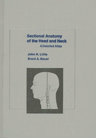
Sectional Anatomy of the Head and Neck: A Detailed Atlas
0
0
As an Amazon Associate and affiliate partner, Menrva Books earns from qualifying purchases. Learn more
Author Description
This superb atlas of the sectional anatomy of the head and neck (exclusive of the CNS) is presented in two complementary planes, coronal and horizontal. Sixty-four exceptional pen-and-ink illustrations accurately capture the fine detail of 3mm thick sections through the region. Every detail was exactly transferred from photographs of the section and its identification was confirmed by microdissection of the exposed surface. These pen-and-ink images have been reproduced at natural size in labeled and unlabeled pairs, an approach that allows the complete identification of structures without masking critical anatomical detail. The captions that accompany each set of illustrations provide brief, authoritative information about the section and how it relates to adjacent tissues. Difficult concepts such as regional compartments and facial spaces are clearly explained, reflecting the broad experience of the senior author in teaching the basic as well as the applied clinical anatomy of the head and neck. Twenty-two sections have been selected for radiographic imaging. These high-resolution radiographs portray planar detail at a level which closely simulates that revealed by high-quality CT images of the region. Introductory chapters review the history of sectional anatomy and provide a unique overview of the fundamental organization of the head and neck. They enhance the value of an atlas that will guide the student and clinician through a sequential study of this anatomically intricate region and will aid radiologists in interpreating bony images and relating them to the soft tissues which surround them.
Menrva's Summary
THE THESIS
This atlas investigates the complex anatomical structures of the head and neck through precise sectional imaging to facilitate clinical understanding. The authors, drawing on extensive experience in teaching applied clinical anatomy, utilize a combination of detailed pen-and-ink illustrations and high-resolution radiographs to map the region. By presenting these sections in coronal and horizontal planes, the text provides a systematic framework for identifying structures and understanding their spatial relationships within the human body.
THE SCOPE MAP
What You Will Find
- Detailed pen-and-ink illustrations of 3mm thick sections of the head and neck.
- Comparative analysis of coronal and horizontal anatomical planes.
- High-resolution radiographic imaging simulating CT scan detail.
- Explanations of complex regional compartments and facial spaces.
THE AUTHORITY PERSPECTIVE
Clinicians and students frequently cite this atlas as a foundational reference for interpreting diagnostic imaging and understanding soft tissue relationships. Experts highlight the clarity of the illustrations and the practical utility of the labeled and unlabeled image pairs for anatomical study.
Page Count:
224
Publication Date:
1994-08-18
Publisher:
Oxford University Press
ISBN-10:
0195042972
ISBN-13:
9780195042979
Science
Medical
Radiology
Neck
Anatomy
Head
Reference
Community Tags
Similar Books