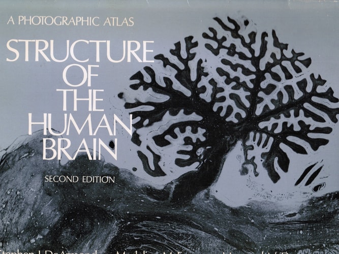
Structure of the Human Brain: A Photographic Atlas
0
0
As an Amazon Associate and affiliate partner, Menrva Books earns from qualifying purchases. Learn more
Author Description
Long awaited, the Third Edition represents a thorough revision of this widely-used atlas. It takes into account the advances in computer-assisted brain-imaging techniques that do not restrict the plane of study and the recent progress in identifying and localizing putative neurotransmitters and neuromodulators in the brain. New sections offer coronal views of the gross brain and brain stem, biochemical neuroanatomy, magnetic resonance images, and recent computerized tomographic scans. In addition, many refinements have been made in the illustrations retained from earlier editions. As before, only the highest quality photomicrographs have been included.
Menrva's Summary
THE THESIS
This atlas investigates the complex anatomical and biochemical architecture of the human brain through high-resolution visual documentation. The authors, experts in neuroanatomy, utilize a combination of traditional photomicrography and modern diagnostic imaging to provide a comprehensive reference for students and practitioners. The text argues that integrating traditional histological study with contemporary computer-assisted imaging is necessary for a complete understanding of brain structure and function.
THE SCOPE MAP
What You Will Find
- Coronal views of the gross brain and brain stem
- Biochemical neuroanatomy and localization of neurotransmitters
- Magnetic resonance imaging (MRI) and computerized tomography (CT) scans
- High-quality photomicrographs of neural tissue
THE AUTHORITY PERSPECTIVE
Experts and medical professionals regard this atlas as a foundational visual resource for neuroanatomical study. Readers frequently note the high technical quality of the imagery and the utility of the updated imaging sections for clinical reference.
Page Count:
208
Publication Date:
1989-04-13
Publisher:
Oxford University Press
ISBN-10:
019504357X
ISBN-13:
9780195043570
Science
Anatomy
Neuroscience
Reference
Medicine
Brain
Community Tags
Similar Books



![Principles of comparative anatomy of invertebrates: [in 2vols]. Vol.2, Organology](https://storage.googleapis.com/menrva_img_storage/covers/generated/9780050021118.jpg)