Radiology Illustrated: Chest Radiology
0
0
As an Amazon Associate and affiliate partner, Menrva Books earns from qualifying purchases. Learn more
Author Description
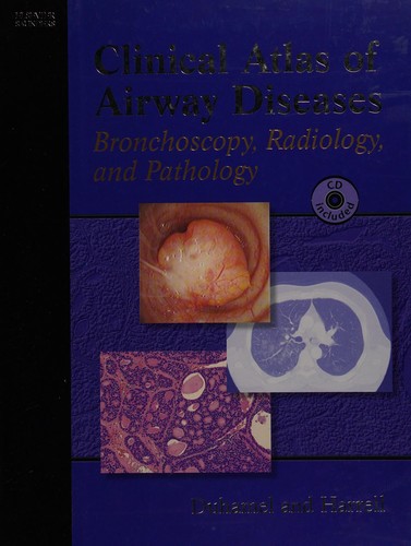
The purpose of this atlas is to illustrate how to achieve reliable diagnoses when confronted by the different abnormalities, or “disease patterns”, that may be visualized on CT scans of the chest. The task of pattern recognition has been greatly facilitated by the advent of high-technology CT such as helical and multidetector CT (MDCT) and dual-energy CT (DECT), and the focus of the book is very much on the role of state-of-the-art MDCT. A wide range of disease patterns and distributions are covered, with emphasis on the typical imaging characteristics of the various focal and diffuse lung diseases. In addition, clinical information relevant to differential diagnosis is provided and the underlying gross and microscopic pathology is depicted, permitting CT–pathology correlation. The entire information relevant to each disease pattern is also tabulated for ease of reference. This book will be an invaluable handy tool that will enable the reader to quickly and easily reach a diagnosis appropriate to the pattern of lung abnormality identified on CT scans. And in this second edition, some more signs and several new diseases and their pattern have been developed and updated. They will be provided with imaging, clinical relevance and CT-pathology correlation. Moreover, the chapters and disease patterns in their order of presentation shall be somewhat changed for readers’ easy legibility and understanding.
Menrva's Summary
THE THESIS
This atlas aims to equip readers with the diagnostic skills to identify abnormalities in chest CT scans by recognizing distinct disease patterns. The book focuses on the application of state-of-the-art multidetector CT (MDCT) and other advanced CT technologies like helical, MDCT, and dual-energy CT (DECT) to facilitate pattern recognition. It covers a broad spectrum of disease patterns and distributions, emphasizing the characteristic imaging features of focal and diffuse lung diseases. Clinical information for differential diagnosis and correlations with underlying gross and microscopic pathology are provided to enhance CT-pathology correlation.
THE SCOPE MAP
- Detailed illustration of disease patterns visualized on chest CT scans.
- Emphasis on the role of state-of-the-art multidetector CT (MDCT).
- Coverage of a wide range of disease patterns and distributions.
- Focus on typical imaging characteristics of focal and diffuse lung diseases.
- Inclusion of clinical information relevant to differential diagnosis.
- Depiction of underlying gross and microscopic pathology for CT-pathology correlation.
- Tabulated information for each disease pattern for ease of reference.
- Updated content in the second edition including new signs, diseases, and patterns.
THE AUTHORITY PERSPECTIVE
The second edition of Radiology Illustrated: Chest Radiology builds upon its predecessor by updating content with new signs and diseases, maintaining its focus on advanced CT technologies like MDCT. The book is structured to facilitate CT-pathology correlation, providing clinical information and pathology details alongside imaging characteristics. Its organization and tabulation of information are designed to serve as a practical tool for readers to quickly reach diagnoses based on identified CT patterns. The revisions aim to enhance legibility and understanding for the reader.
Page Count:
900
Publication Date:
2024-02-09
Publisher:
Springer Nature
ISBN-10:
9819966337
ISBN-13:
9789819966332
Science & Technology
Non-Fiction
Pathology
Radiology
Pulmonology
Community Tags
Similar Books