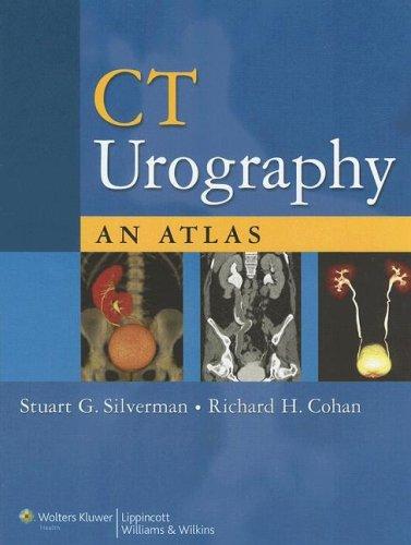
CT Urography
0
0
As an Amazon Associate and affiliate partner, Menrva Books earns from qualifying purchases. Learn more
Author Description
Featuring over 500 images, this atlas is the first text on performing and interpreting CT urography. Chapters detail the indications and techniques for CT urography, review the risks of radiation exposure, show how normal urinary tract anatomy and variants appear on CT scans, and demonstrate a wide range of urinary tract abnormalities as they appear on thin-section CT. The final chapter illustrates artifacts and diagnostic pitfalls. Chapters on abnormalities follow a case-based teaching file format. Each case is presented on a two-page spread, with images and succinct discussion of the entity and how CT urography was used to diagnose it.
Menrva's Summary
THE THESIS
This atlas serves as the definitive guide to the practice and interpretation of CT urography. It meticulously covers the indications and techniques for performing the procedure, addresses concerns regarding radiation exposure, and provides detailed anatomical references for normal urinary tract variations as visualized on CT scans. The core of the atlas is dedicated to showcasing a broad spectrum of urinary tract abnormalities through thin-section CT imaging, presented in a case-based teaching file format.
THE SCOPE MAP
- Detailed coverage of indications and techniques for CT urography.
- Discussion on risks associated with radiation exposure.
- Comprehensive review of normal urinary tract anatomy and variants on CT.
- Extensive demonstration of urinary tract abnormalities using thin-section CT.
- Case-based teaching file format for abnormalities.
- Two-page spread for each case, featuring images and concise diagnostic discussions.
- Identification of artifacts and diagnostic pitfalls.
THE AUTHORITY PERSPECTIVE
This comprehensive atlas is recognized as a foundational text for radiologists and urologists seeking to master CT urography. Its detailed imaging and case-based approach are designed to equip practitioners with the skills necessary for accurate interpretation and diagnosis of a wide array of urinary tract conditions. The inclusion of radiation safety information and common pitfalls further enhances its utility as a practical reference.
Page Count:
272
Publication Date:
2006-01-01
ISBN-10:
0781787548
ISBN-13:
9780781787543
Non-Fiction
Science
Atlas
Radiology
Urology
textbook
Community Tags
Similar Books