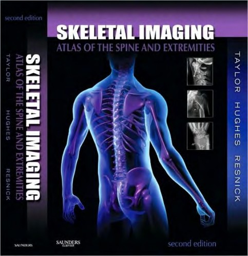
Skeletal Imaging
0
0
As an Amazon Associate and affiliate partner, Menrva Books earns from qualifying purchases. Learn more
Author Description
Use this atlas to accurately interpret images of musculoskeletal disorders! Taylor, Hughes, and Resnick₂s Skeletal Imaging: Atlas of the Spine and Extremities, 2nd Edition covers each anatomic region separately, so common disorders are shown within the context of each region. This allows you to examine and compare images for a variety of different disorders. A separate chapter is devoted to each body region, with coverage of normal developmental anatomy, developmental anomalies and normal variations, and how to avoid a misdiagnosis by differentiating between disorders that appear to be similar. All of the most frequently encountered musculoskeletal conditions are included, from physical injuries to tumors to infectious diseases. Over 2,100 images include radiographs, radionuclide studies, CT scans, and MR images, illustrating pathologies and comparing them with other disorders in the same region. Organization by anatomic region addresses common afflictions for each region in separate chapters, so you can see how a particular region looks when affected by one condition as compared to its appearance with other conditions. Coverage of each body region includes normal developmental anatomy, fractures, deformities, dislocations, infections, hematologic disorders, and more. Normal Developmental Anatomy sections open each chapter, describing important developmental landmarks in various regions of the body from birth to skeletal maturity. Practical tables provide a quick reference to essential information, including normal developmental anatomic milestones, developmental anomalies, common presentations and symptoms of diseases, and much more.
Menrva's Summary
THE THESIS
This atlas provides a comprehensive guide to the interpretation of skeletal imaging, focusing on musculoskeletal disorders of the spine and extremities. The book is organized by anatomic region, allowing for direct comparison of various pathologies within their regional context. It covers normal developmental anatomy, anomalies, and variations, alongside common conditions such as physical injuries, tumors, and infectious diseases, aiming to prevent misdiagnosis by differentiating similar-appearing disorders. The text utilizes over 2,100 images, including radiographs, radionuclide studies, CT scans, and MR images, to illustrate pathologies and aid in comparative analysis.
THE SCOPE MAP
- Coverage organized by distinct anatomic regions of the spine and extremities.
- Detailed examination of normal developmental anatomy for each region.
- Inclusion of developmental anomalies and normal variations.
- Focus on differentiating between disorders that present with similar imaging characteristics.
- Comprehensive coverage of frequently encountered musculoskeletal conditions, including physical injuries, tumors, and infectious diseases.
- Over 2,100 images featuring radiographs, radionuclide studies, CT scans, and MR images.
- Practical tables for quick reference on developmental milestones, anomalies, and disease presentations.
- Emphasis on avoiding misdiagnosis through comparative imaging analysis.
THE AUTHORITY PERSPECTIVE
This atlas is structured to facilitate accurate interpretation of skeletal imaging, particularly for musculoskeletal disorders. The organization by anatomic region allows for direct comparison of pathologies, aiding in differential diagnosis. Coverage includes normal anatomy, developmental variations, and a wide array of common conditions, supported by an extensive collection of imaging studies. The inclusion of practical tables further enhances its utility as a reference tool for clinicians and students.
Page Count:
0
Publication Date:
2010-01-01
ISBN-10:
1416056238
ISBN-13:
9781416056232
Science
Non-Fiction
Orthopedics
Radiology
Anatomy
Community Tags
Similar Books