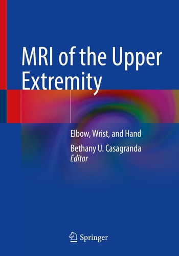
MRI of the Upper Extremity: Elbow, Wrist, and Hand
0
0
As an Amazon Associate and affiliate partner, Menrva Books earns from qualifying purchases. Learn more
Author Description
This book systematically discusses the anatomy and pathology of three specific regions of the upper extremity: the elbow, wrist, and hand. Divided into three sections, by body part, chapters cover anatomy and pathology. The anatomy chapters give a comprehensive view of each body part and normal variants found there. Although the primary modality emphasized will be MRI, illustrations and other modalities, including plain radiograph and CT, will be used to comprehensively discuss the anatomy of each region. Liberally illustrated, the pathology chapters then cover both traumatic and non-traumatic causes for imaging and detail how to perform and interpret each MRI. Specific examples include: osseous trauma, soft tissue trauma, and tumor imaging. Chapters are written with the deliberate intention to be of value to all levels of radiology training while remaining a reliable resource for attending radiologists.
Menrva's Summary
THE THESIS
This book provides a systematic exploration of the anatomy and pathology of the elbow, wrist, and hand, focusing primarily on Magnetic Resonance Imaging (MRI). It is divided into three distinct sections, each dedicated to a specific body part, with chapters covering both normal anatomy and pathological conditions. The anatomy chapters offer a thorough overview, incorporating plain radiographs and CT scans alongside MRI to illustrate normal variants and structures. The pathology chapters detail the imaging approach and interpretation for both traumatic and non-traumatic conditions, including osseous trauma, soft tissue injuries, and tumor imaging, aiming to serve radiologists at all training levels.
THE SCOPE MAP
- Detailed anatomical review of the elbow, wrist, and hand.
- Comprehensive coverage of normal variants across imaging modalities.
- Pathology chapters focusing on traumatic injuries of the elbow, wrist, and hand.
- Pathology chapters addressing non-traumatic conditions, including tumors.
- Guidance on performing and interpreting MRI scans for upper extremity pathologies.
- Integration of plain radiograph and CT illustrations to supplement MRI.
- Content structured to benefit both trainees and practicing radiologists.
- Specific examples of osseous trauma, soft tissue trauma, and tumor imaging.
THE AUTHORITY PERSPECTIVE
The book is structured to be a valuable resource for radiologists at all levels, from trainees to attending physicians. Its systematic approach, dividing content by body part (elbow, wrist, hand), ensures comprehensive coverage of both normal anatomy and a wide range of pathologies. By integrating MRI with other modalities like plain radiography and CT, it aims to provide a thorough understanding of the upper extremity. The inclusion of specific examples such as osseous trauma, soft tissue trauma, and tumor imaging further enhances its practical utility for diagnostic interpretation.
Page Count:
279
Publication Date:
2021-10-10
Publisher:
Springer International Publishing
ISBN-10:
3030816117
ISBN-13:
9783030816117
Non-Fiction
Science
Orthopedics
Anatomy
Pathology
Radiology
Community Tags
Similar Books