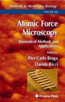
Atomic Force Microscopy
0
0
As an Amazon Associate and affiliate partner, Menrva Books earns from qualifying purchases. Learn more
Author Description
Provides biomedical researchers the hands-on knowledge for using atomic force microscopy in their research. It offers many benefits including high manification with high resolution, minimal tissue or cell preparation, the ability to obtain different views of the sample from a single data collection, and the ability to study live cells in their physiologic environment. The authors begin with an introduction on the basics of atomic force microscopy and then covers specific protocols for the morpho-structural analysis of cellular structures. Also covered are detailed protocols for investigating subcellular structures and the latest techniques for functional investigations using AFM. The 28 chapters should prove to be an essential laboratory manual for all scientists using atomic force microscopy in their biomedical research.
Menrva's Summary
THE THESIS
This manual equips biomedical researchers with the practical knowledge to implement atomic force microscopy (AFM) in their studies. It details the advantages of AFM, such as its high magnification and resolution, minimal sample preparation, versatile imaging capabilities, and the unique ability to observe live cells in their natural state. The text progresses from fundamental AFM principles to specific protocols for analyzing cellular and subcellular structures, concluding with advanced techniques for functional investigations.
THE SCOPE MAP
- Introduction to the fundamental principles of Atomic Force Microscopy (AFM).
- Protocols for morpho-structural analysis of cellular structures.
- Detailed protocols for investigating subcellular structures.
- Latest techniques for functional investigations using AFM.
- Applications of AFM in biomedical research.
- High magnification and resolution imaging.
- Minimal tissue or cell preparation requirements.
- Study of live cells in their physiologic environment.
THE AUTHORITY PERSPECTIVE
This book is presented as an essential laboratory manual for scientists engaged in biomedical research utilizing atomic force microscopy. The comprehensive nature of its 28 chapters, covering basic principles through advanced functional investigations, suggests it aims to be a thorough resource. The focus on practical, hands-on knowledge indicates its utility for researchers seeking to apply AFM techniques directly to their work.
Page Count:
394
Publication Date:
2004-01-01
ISBN-10:
1588290948
Science
Non-Fiction
Biomedical Research
Cell Biology
Microscopy
Laboratory Manual
Community Tags
Similar Books