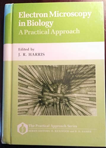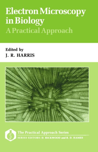
Electron Microscopy in Biology: A Practical Approach
0
0
As an Amazon Associate and affiliate partner, Menrva Books earns from qualifying purchases. Learn more
Author Description
This volume provides detailed protocols for a wide range of procedures relating to the electron microscopy of tissues, cells, and their components with the emphasis on biomedical applications. The fundamental methods for specimen preparation, tissue fixation, embedding, and sectioning are covered in depth and are followed by techniques in freeze-substitution, immunogold labelling, auto- radiography, and enzyme histochemistry. Although the technical details in the book relate mainly to tissues and cells, many of the procedures can be adapted for isolated organelles and membrane fractions, viral particles, macromolecular assemblies, and large proteins. This is a broadly based compilation of the most important specimen preparation techniques and their applications. It will be of interest to researchers and advanced students working in almost every biomedical discipline, plus technicians in electron microscopy units.
Menrva's Summary
THE THESIS
This volume investigates the standardized protocols and technical methodologies required for effective electron microscopy in biological and biomedical research. Robin Harris compiles a comprehensive set of procedures designed to assist researchers and laboratory technicians in navigating the complexities of specimen preparation. By focusing on the intersection of tissue analysis and imaging, the text provides a structured framework for achieving high-quality results in diverse biological applications.
THE SCOPE MAP
What You Will Find
- Detailed protocols for tissue fixation, embedding, and sectioning techniques.
- Advanced methodologies including freeze-substitution and immunogold labelling.
- Specialized applications for enzyme histochemistry and autoradiography.
- Adaptable procedures for isolated organelles, viral particles, and macromolecular assemblies.
THE AUTHORITY PERSPECTIVE
Experts recognize this volume as a reliable technical reference for laboratory settings, particularly for those performing routine biomedical imaging. Readers frequently note the practical utility of the step-by-step instructions provided for complex specimen preparation.
Page Count:
328
Publication Date:
1991-12-26
Publisher:
IRL Press
ISBN-10:
0199632154
ISBN-13:
9780199632152
Science
Biology
Laboratory Manual
Microscopy
Biomedical Research