Atlas of Electromyography
0
0
As an Amazon Associate and affiliate partner, Menrva Books earns from qualifying purchases. Learn more
Author Description
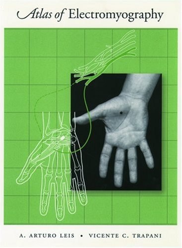
The Atlas of Electromyography is a visually alluring book which provides high quality anatomical illustrations of skeletal muscles that include nerve, plexus, and root supply; photographs of each muscle in healthy subjects to enable the practitioner to identify the optimum site of EMG needle insertion; clinical features of the major conditions affecting peripheral nerves; and electrodiagnostic strategies for confirming suspected lesions of the peripheral nervous system. The atlas is divided into sections on the major peripheral nerves. Each nerve is illustrated and its anatomy reviewed in the text. The authors provide a detailed outline of the clinical conditions and entrapment syndromes that affect the nerve, including a list of etiologies, clinical features, and electrodiagnostic strategies used for each symdrome. Each muscle supplied by the peripheral nerve is shown as an anatomical illustration with a corresponding human photograph. The text provides information about the muscle origin, tendon insertion, voluntary activation maneuver, and site of optimum needle insertion. The needle insertion point is identified in both the anatomical illustration and the corresponding photographs. This assures that pertinent bone, muscular, and soft tissue landmarks can be used to guide the electromyographer to a specific point on the skin. Potential pitfalls associated with the needle insertion are added, usually noting adjacent muscles or structures that may be mistakenly entered. Clinical correlates pertinent to the muscle being examined are also provided. The tlas of Electromyography serves as an anatomical guide for practitioners of electromyography and neurologists, as well as residents i neurology, physical medicine, and rehabilitation.
Menrva's Summary
THE THESIS
This text serves as a comprehensive visual and clinical guide designed to improve the accuracy and safety of electromyography (EMG) needle insertion procedures. The authors, A. Arturo Leis and Vicente C. Trapani, utilize a combination of detailed anatomical illustrations and clinical photography to bridge the gap between theoretical nerve anatomy and practical bedside application. By providing specific landmarks and diagnostic strategies, the work establishes a standardized framework for identifying peripheral nerve lesions and entrapment syndromes.
THE SCOPE MAP
What You Will Find
- Detailed anatomical illustrations of skeletal muscles including nerve, plexus, and root supply.
- High-quality photographs of healthy subjects demonstrating optimal needle insertion sites.
- Comprehensive outlines of clinical conditions and entrapment syndromes affecting peripheral nerves.
- Practical guidance on identifying bone, muscular, and soft tissue landmarks to avoid potential procedural pitfalls.
THE AUTHORITY PERSPECTIVE
Practitioners and residents in neurology and physical medicine frequently cite this atlas as a foundational resource for mastering the technical aspects of electrodiagnostic testing. Experts highlight the clarity of the visual aids and the clinical relevance of the provided anatomical correlates as essential for daily practice.
Page Count:
199
Publication Date:
2000-01-01
Publisher:
Oxford University Press
ISBN-10:
0198027222
ISBN-13:
9780198027225
Anatomy
Reference
Medical
Allied Health
Neurology