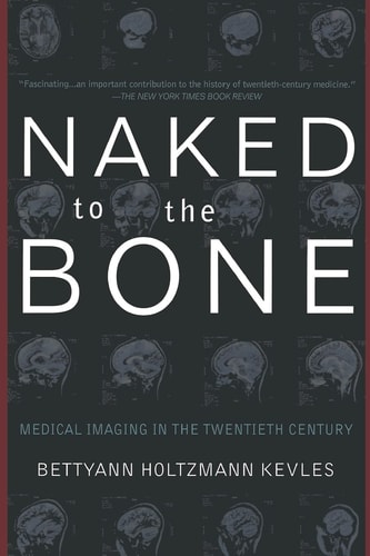
Naked to the Bone: Medical Imaging in the Twentieth Century
Author Description
A century ago, the living body, like most of the material world, was opaque. Then Wilhelm Roentgen captured and X-ray image of his wife's finger—her wedding ring “floating” around a white bone—and our range of vision changed forever. By the 1920s, X-ray technology was common-place: all army recruits had lined up for chest pictures during WWI, and children were examining the bones of their feet in shoe store fluoroscopes, spectacularly unaware of the radiation they were absorbing. Through lucid prose, vivid anecdotes, and over seventy striking illustrations, science writer Bettyann Holtzman Kevles shows how X-rays and the subsequent daughter technologies—CT, MRI, PET, ultrasound—transformed the practice of medicine (from pediatrics to neurosurgery), the rules of evidence in courts, and the vision of artists.
Menrva's Summary
THE THESIS
This book investigates how the development and proliferation of medical imaging technologies fundamentally altered human perception of the body, medical practice, and legal standards throughout the twentieth century. Author Bettyann Holtzmann Kevles, a science historian, utilizes a combination of archival research, historical anecdotes, and technical analysis to trace the evolution of imaging from the discovery of X-rays to the advent of MRI and PET scans. She argues that these tools did more than improve diagnostics; they redefined the boundaries of privacy and the nature of physical evidence in modern society.
THE SCOPE MAP
What You Will Find
- Historical progression of imaging technologies including X-rays, CT scans, MRI, PET, and ultrasound.
- Analysis of the cultural impact of medical imaging on art, law, and public health.
- Case studies detailing the integration of imaging into diverse medical fields such as pediatrics and neurosurgery.
- Examination of the ethical and safety implications regarding radiation exposure and diagnostic reliance.
THE AUTHORITY PERSPECTIVE
Experts and historians frequently cite this work as a comprehensive and accessible survey of the intersection between medical technology and twentieth-century culture. Readers often note the clarity of the prose, which successfully bridges the gap between complex technical history and its broader societal consequences.
Page Count:
378
Publication Date:
1998-03-19
Publisher:
Basic Books
ISBN-10:
020132833X
ISBN-13:
9780201328332
Non-Fiction
History
Science
Technology
Medicine
Community Tags
Similar Books