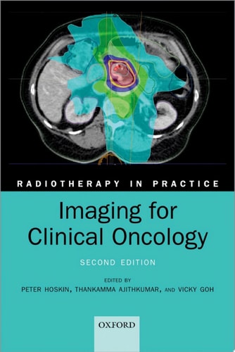
Imaging for Clinical Oncology
0
0
As an Amazon Associate and affiliate partner, Menrva Books earns from qualifying purchases. Learn more
Author Description
Imaging is a critical component in the delivery of radiotherapy to patients with malignancy, and this book teaches the principles and practice of imaging specific to radiotherapy. Introductory chapters outline the basic principles of the available imaging modalities including x-rays, CT, ultrasound, MRI, nuclear medicine, and PET. Site specific chapters then cover the main tumour sites, reviewing optimal imaging techniques for diagnosis, staging, radiotherapy planning, and follow-up for each site. The important areas of radiation protection, exposure justification, and risks are also covered, exploring issues such as balancing radiation exposure with long-term risks of radiation effects, such as second cancer induction. This second edition has been fully revised and updated to reflect current techniques, and includes two brand new chapters on imaging for radiotherapy treatment verification, and the role of specialist MRI techniques and functional imaging for radiotherapy planning. With insights from experts in each field and over 200 illustrations, this comprehensive and easy-to-read guide will be an invaluable resource for radiation oncologists, clinical oncologists, and radiotherapists, both qualified and in training. ABOUT THE SERIES Radiotherapy remains the major non-surgical treatment modality for the management of malignant disease. It is based on the application of the principles of applied physics, radiobiology, and tumour biology to clinical practice. Each volume in the series takes the reader through the basic principles of the use of ionizing radiation and then develops this by individual sites. This series of practical handbooks is aimed at physicians both training and practising in radiotherapy, as well as medical physics, dosimetrists, radiographers, and senior nurses.
Menrva's Summary
THE THESIS
This text investigates the integration of advanced imaging modalities into the clinical workflow of radiotherapy for malignant disease. Author Daniel Ogden provides a technical framework for radiation oncologists and radiotherapists, synthesizing principles of physics and clinical oncology to optimize patient outcomes. The book argues that precise imaging is fundamental to accurate diagnosis, staging, treatment planning, and long-term follow-up in cancer care.
THE SCOPE MAP
What You Will Find
- Technical overviews of X-ray, CT, ultrasound, MRI, nuclear medicine, and PET modalities.
- Site-specific protocols for tumor diagnosis and radiotherapy planning.
- Analysis of radiation protection, exposure justification, and risk management.
- Updated chapters on treatment verification and functional MRI techniques.
Scope Limits
- Does not provide exhaustive surgical or chemotherapy protocols.
- Focuses primarily on radiotherapy applications rather than general diagnostic radiology.
THE AUTHORITY PERSPECTIVE
Experts and practitioners frequently cite this volume as a foundational handbook for those specializing in radiation oncology. Readers note the text's balance between theoretical physics and practical clinical application, making it a standard reference for both trainees and established professionals.
Page Count:
393
Publication Date:
2021-01-01
Publisher:
OUP Oxford
ISBN-10:
0192637258
ISBN-13:
9780192637253
Radiotherapy
Allied Health
Medical Physics
Medical
Oncology
Radiology
Community Tags
Similar Books